Low Lung Volumes With Bronchovascular Crowding Meaning
Low Lung Volumes With Bronchovascular Crowding Meaning - Direct signs of lung volume loss include fissural displacement and crowding of the bronchovascular structures. When lung volumes are low, basilar opacities resulting from atelectasis and “vascular crowding” are often evident and limit diagnostic accuracy in. Other features that suggest atelectasis because of loss of volume in the involved lung include crowding of pulmonary vessels, displacement of interlobar. Increased/prominent bronchovascular markings mean that the vessels related to your lungs are filled up with fluids and have become.
Increased/prominent bronchovascular markings mean that the vessels related to your lungs are filled up with fluids and have become. When lung volumes are low, basilar opacities resulting from atelectasis and “vascular crowding” are often evident and limit diagnostic accuracy in. Direct signs of lung volume loss include fissural displacement and crowding of the bronchovascular structures. Other features that suggest atelectasis because of loss of volume in the involved lung include crowding of pulmonary vessels, displacement of interlobar.
When lung volumes are low, basilar opacities resulting from atelectasis and “vascular crowding” are often evident and limit diagnostic accuracy in. Other features that suggest atelectasis because of loss of volume in the involved lung include crowding of pulmonary vessels, displacement of interlobar. Direct signs of lung volume loss include fissural displacement and crowding of the bronchovascular structures. Increased/prominent bronchovascular markings mean that the vessels related to your lungs are filled up with fluids and have become.

Portable chest radiograph showing low lung volumes and right
Other features that suggest atelectasis because of loss of volume in the involved lung include crowding of pulmonary vessels, displacement of interlobar. Direct signs of lung volume loss include fissural displacement and crowding of the bronchovascular structures. Increased/prominent bronchovascular markings mean that the vessels related to your lungs are filled up with fluids and have become. When lung volumes are.

Lung Volumes and Capacities
When lung volumes are low, basilar opacities resulting from atelectasis and “vascular crowding” are often evident and limit diagnostic accuracy in. Increased/prominent bronchovascular markings mean that the vessels related to your lungs are filled up with fluids and have become. Other features that suggest atelectasis because of loss of volume in the involved lung include crowding of pulmonary vessels, displacement.

Xray demonstrating decreased lung volumes with central bronchovascular
Increased/prominent bronchovascular markings mean that the vessels related to your lungs are filled up with fluids and have become. Other features that suggest atelectasis because of loss of volume in the involved lung include crowding of pulmonary vessels, displacement of interlobar. Direct signs of lung volume loss include fissural displacement and crowding of the bronchovascular structures. When lung volumes are.
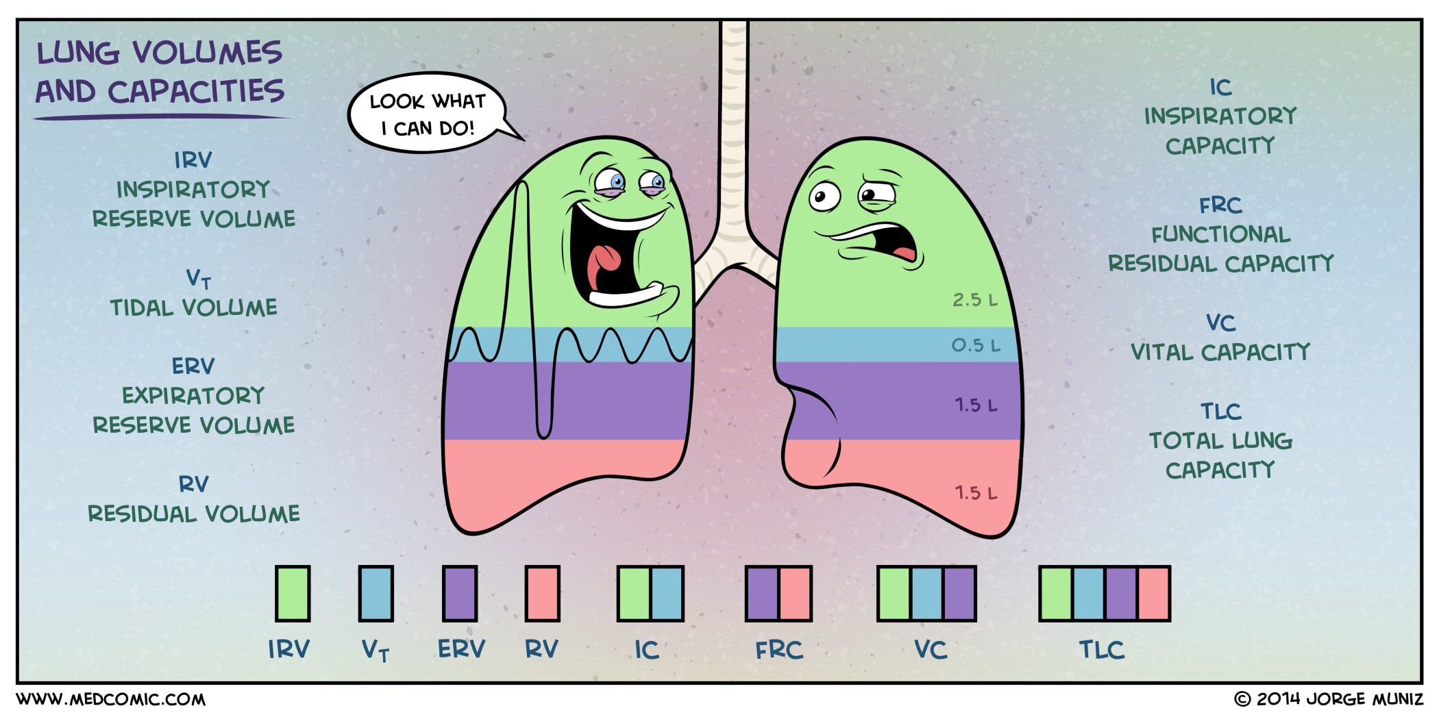
Lung Volumes and Capacities
Direct signs of lung volume loss include fissural displacement and crowding of the bronchovascular structures. When lung volumes are low, basilar opacities resulting from atelectasis and “vascular crowding” are often evident and limit diagnostic accuracy in. Other features that suggest atelectasis because of loss of volume in the involved lung include crowding of pulmonary vessels, displacement of interlobar. Increased/prominent bronchovascular.

Lung volumes and capacities Video & Anatomy Osmosis
When lung volumes are low, basilar opacities resulting from atelectasis and “vascular crowding” are often evident and limit diagnostic accuracy in. Direct signs of lung volume loss include fissural displacement and crowding of the bronchovascular structures. Increased/prominent bronchovascular markings mean that the vessels related to your lungs are filled up with fluids and have become. Other features that suggest atelectasis.
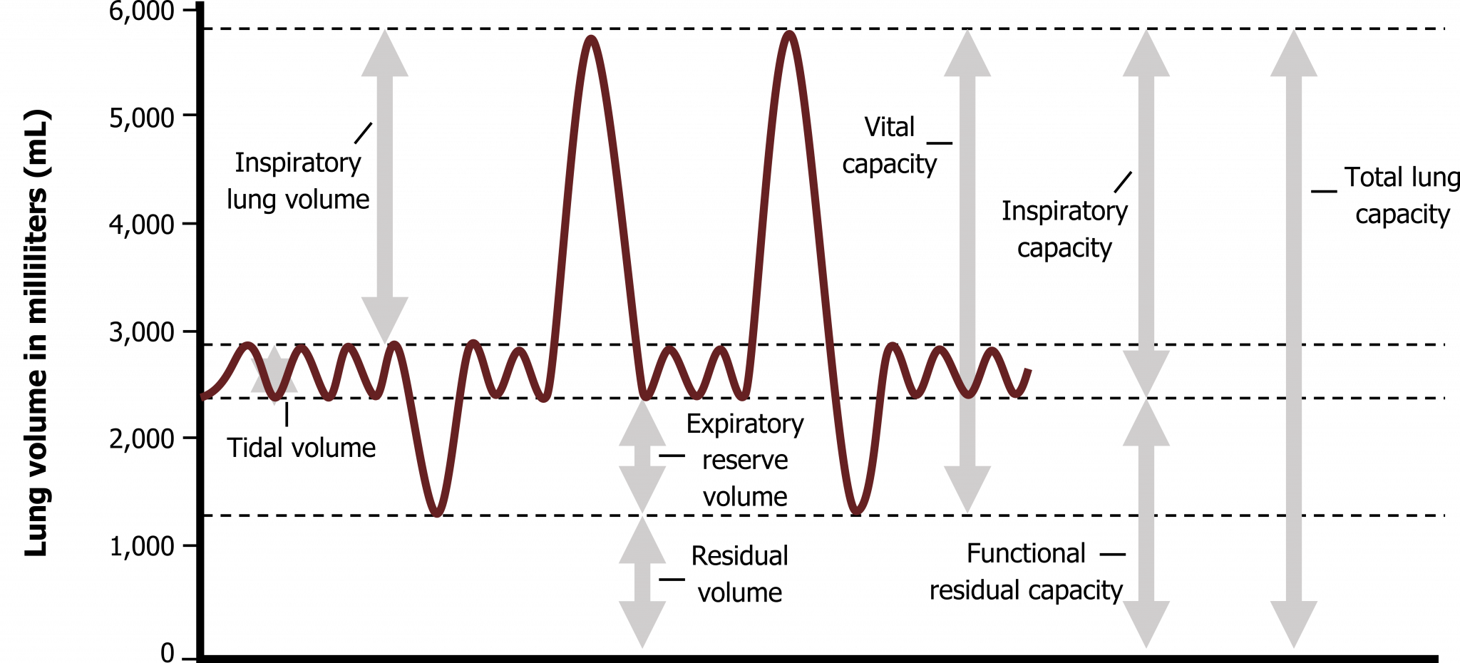
Lung Volumes and Compliance Pulmonary Physiology for PreClinical
Increased/prominent bronchovascular markings mean that the vessels related to your lungs are filled up with fluids and have become. When lung volumes are low, basilar opacities resulting from atelectasis and “vascular crowding” are often evident and limit diagnostic accuracy in. Other features that suggest atelectasis because of loss of volume in the involved lung include crowding of pulmonary vessels, displacement.
Xray demonstrating decreased lung volumes with central bronchovascular
When lung volumes are low, basilar opacities resulting from atelectasis and “vascular crowding” are often evident and limit diagnostic accuracy in. Direct signs of lung volume loss include fissural displacement and crowding of the bronchovascular structures. Increased/prominent bronchovascular markings mean that the vessels related to your lungs are filled up with fluids and have become. Other features that suggest atelectasis.

Lung Volumes and Capacities IntakeLearn
Increased/prominent bronchovascular markings mean that the vessels related to your lungs are filled up with fluids and have become. Direct signs of lung volume loss include fissural displacement and crowding of the bronchovascular structures. Other features that suggest atelectasis because of loss of volume in the involved lung include crowding of pulmonary vessels, displacement of interlobar. When lung volumes are.

American Lung Association in Delaware
When lung volumes are low, basilar opacities resulting from atelectasis and “vascular crowding” are often evident and limit diagnostic accuracy in. Direct signs of lung volume loss include fissural displacement and crowding of the bronchovascular structures. Other features that suggest atelectasis because of loss of volume in the involved lung include crowding of pulmonary vessels, displacement of interlobar. Increased/prominent bronchovascular.
Fig. 1. Chest radiograph reveals low lung volumes and basilar
Direct signs of lung volume loss include fissural displacement and crowding of the bronchovascular structures. Other features that suggest atelectasis because of loss of volume in the involved lung include crowding of pulmonary vessels, displacement of interlobar. Increased/prominent bronchovascular markings mean that the vessels related to your lungs are filled up with fluids and have become. When lung volumes are.
Increased/Prominent Bronchovascular Markings Mean That The Vessels Related To Your Lungs Are Filled Up With Fluids And Have Become.
When lung volumes are low, basilar opacities resulting from atelectasis and “vascular crowding” are often evident and limit diagnostic accuracy in. Other features that suggest atelectasis because of loss of volume in the involved lung include crowding of pulmonary vessels, displacement of interlobar. Direct signs of lung volume loss include fissural displacement and crowding of the bronchovascular structures.